Custom-built Raman Microspectroscopy

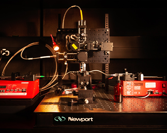
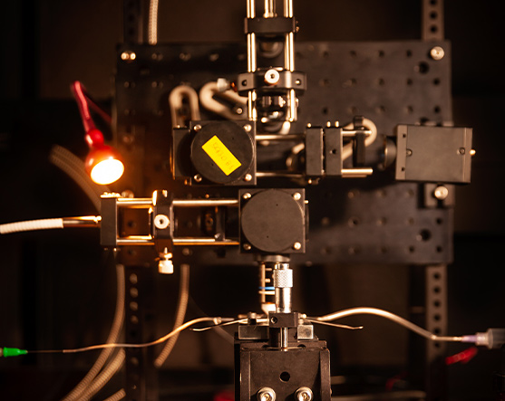
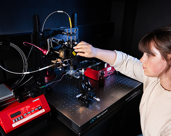
This is a compact Raman micro-spectroscopy setup that is completely customizable in the spirit of “Lego-Optics”. Laser spot engineering enables laser spot diameter at the sample focus ranging from 1-30 μm. The depth of focus can be adjusted from ~ 1 μm (for confocal Raman spectroscopy) to 10-100 μm (for flow-cell Raman spectroscopy of liquid samples). Snapshot depth-sensitive Raman spectroscopy (for layered samples such as radiochromic films) is possible using a Bessel beam profile (coming soon) of the excitation 785 nm laser. By means of raster scanning or point-by-point scanning of the microscope stage, this setup enables label-free chemical mapping of specimens for rapid molecular fingerprinting as a function of spatial location.

Technical Specifications

  • Choice of two input lasers at 785 nm that are fiber-coupled: 100 mW, single-mode CW laser (IPS) for 1 mm beam spot diameter, and 500 mW multi-mode CW laser (Ondax) for 10-30 mm beam spot diameter.
  • Andor Spectrometer with 4 cm-1 spectral resolution
  • Sample format: Liquid or Dry biological cells (live or fixed), solid and liquid tissue such as blood.
  • Microscope objectives for excitation and collection: 60X (1.1 NA) water immersion, 40X (0.75 NA) water immersion
  • Raman spectroscopic mapping with motorized or piezo-driven scanning stages

Interested in
learning more?

We always look forward to hearing from motivated potential collaborators, partners, and trainees.

Contact Us Newsletter Form

This site is protected by reCAPTCHA and the Google Privacy Policy and Terms of Service apply.

Interested in Exploring More?

The Team

We are a group of experts from different disciplines working together to tackle one of the biggest challenges of modern times: the characterization and recreation of tissue microenvironments.

Research

We seek answers to questions related to tissue homeostasis and disease. New technologies resulting from our work will facilitate diagnosis and treatment of many chronic diseases.

Infrastructure

Our state-of-the-art microscopy and analytical techniques allow us to analyze the complex biochemical and biophysical processes that occur in tissue microenvironments.

Training

By providing research opportunities and developing educational programs for the larger community, we are helping Ontario and, indeed, all of Canada build expertise in emerging but increasingly important areas of research, including tissue engineering and imaging.