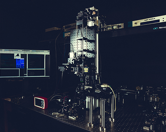
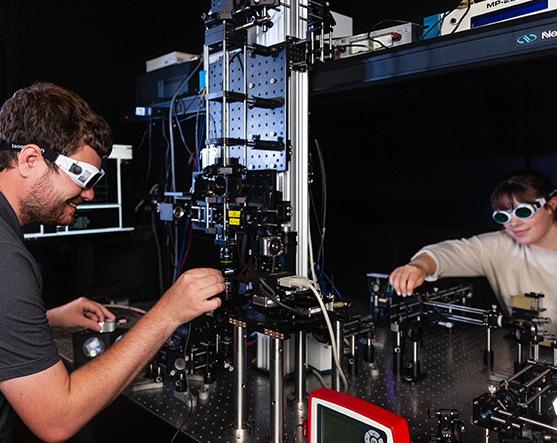
This is an open frame, custom-configurable laser-scanning microscope coupled to a dual output ~100 femtosecond pulsed laser for coherent Raman scattering and Nonlinear Optical Microscopy (NLOM) imaging. One of the laser outputs, the “Pump beam” can be optically switched between one of two paths such that i) the 100 fs pulses are used for Two Photon Excitation Fluorescence (TPEF), and Second Harmonic Generation (SHG) imaging or ii) ~2 ps pulses are generated (by spectral focusing) for coherent Raman imaging e.g. Coherent Anti-Stokes Raman Scattering (CARS) and Stimulated Raman Scattering (SRS) imaging. The TPEF and SHG imaging enables characterization of e.g. the metabolic activity in mitochondria and the remodeling of the collagen respectively. The CARS and SRS imaging in the CH-stretch region of 2800 – 3050 cm-1 of Raman shifts is useful for label-free mapping of the changes in lipids (2855 cm-1), proteins (2930 cm-1) and DNA (2955 cm-1).


Technical Specifications
- Insight DS+ femtosecond pulsed laser (MKS-Spectra Physics) with two synchronized free-space outputs of ~ 100 fs and 220 fs pulses emitted at a repetition rate of 80 MHz.
- One output can be tuned in the 680 – 1300 nm range with >1 W average power
- The second output is fixed at 1040 nm range with >500 mW average power.
- Chirped pulses up to 2.5 ps generated for higher spectral resolution CARS and SRS imaging.
- Upright laser-scanning microscope utilizing galvo-galvo scanning
- Epi-TPEF, forward-CARS, SRS and SHG imaging
- Microscope objectives: 60X (1.1 NA) water immersion, 20X air (0.8 NA)
- Signal Collection optics: 40X (0.75 NA) water immersion
ScanImage open-source software based on Matlab for laser scanning and image generation
Interested in
learning more?
We always look forward to hearing from motivated potential collaborators, partners, and trainees.
Contact Us Newsletter Form
Interested in Exploring More?
The Team
We are a group of experts from different disciplines working together to tackle one of the biggest challenges of modern times: the characterization and recreation of tissue microenvironments.
Research
We seek answers to questions related to tissue homeostasis and disease. New technologies resulting from our work will facilitate diagnosis and treatment of many chronic diseases.
Infrastructure
Our state-of-the-art microscopy and analytical techniques allow us to analyze the complex biochemical and biophysical processes that occur in tissue microenvironments.
Training
By providing research opportunities and developing educational programs for the larger community, we are helping Ontario and, indeed, all of Canada build expertise in emerging but increasingly important areas of research, including tissue engineering and imaging.