
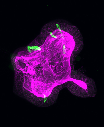
Mitochondria are the metabolic hub of the cell. During bacterial and viral infections, mitochondrial function is compromised leading to unregulated immune responses of the hosts’ cells. In this study, we investigated mitochondrial dysfunction in murine cells stimulated with bacterial (LPS) and viral (Poly(I:C)) ligands. We utilized label-free two-photon excitation fluorescence (TPEF) imaging of endogenous fluorophores, reduced nicotinamide adenine dinucleotide phosphate (NAD(P)H) and flavin adenine dinucleotide (FAD) levels. In addition to evaluating mitochondrial function utilizing a standard metric such as the optical redox ratio, we developed a new metric for the first time based on the FAD-TPEF intensity to quantify mitochondrial organization. Our results show that TPEF imaging is a robust label-free method to examine cellular mitochondrial activity in immune cells such as macrophages.
Publication
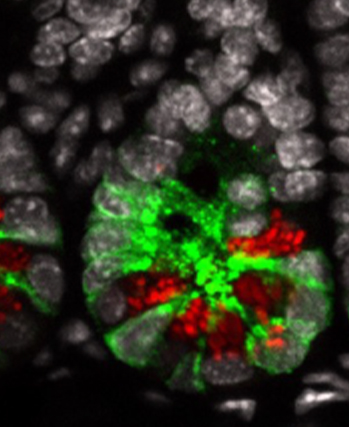
BMP signaling in the intestinal epithelium drives a critical feedback loop to restrain IL-13-driven tuft cell hyperplasia
Although Helminth infections are prevalent throughout the world, they are particular a health...
Publication
LSD1 represses a neonatal/reparative gene program in adult intestinal epithelium
After birth, the epithelium that lines our gut transitions (or matures) so that it can deal...
Publication
Targeting mRNA binding proteins to respond to stress responses
The paper establishes the phosphorylation of heterogeneous nuclear ribonucleoprotein A1...
